A Clinical Perspective for Indian Pulmonologists, Chest Physicians, and Respiratory Specialists
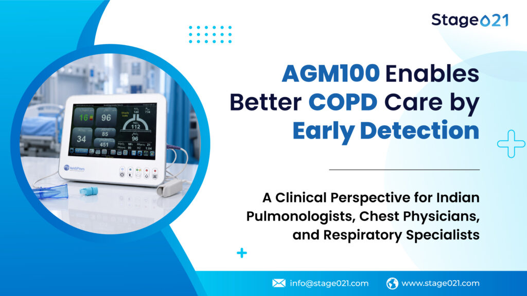
India's COPD Crisis: A Scale That Demands Rethinking
India carries a respiratory burden that is, by any measure, extraordinary. According to the Global Burden of Disease (GBD) 2019 report, there are an estimated 37.8 million people living with COPD in India, representing 17.8% of the global COPD burden. More strikingly, India contributes a disproportionate 27.3% of global COPD deaths, despite having roughly 18% of the world’s population. COPD is now the second leading cause of death and disability-adjusted life years (DALYs) in India, a ranking that places it above stroke, diabetes, and most infectious diseases in terms of population-level disease burden.
And yet, for a disease of this magnitude, India’s diagnostic infrastructure is profoundly mismatched to its clinical reality. Spirometry, the current gold standard for COPD diagnosis, is underused, unavailable in most primary care settings, inaccessible to rural populations, and structurally biased toward detecting disease only after significant irreversible lung damage has already occurred.
India’s COPD story is not merely a story of high prevalence. It is a story of a disease that is simultaneously widespread, underdiagnosed, under-detected before symptoms, and clinically misunderstood in its full physiological complexity. For Indian clinicians, this creates both a profound diagnostic challenge and, with new technology, an unprecedented clinical opportunity.
This article makes the case that gas exchange assessment, specifically the non-invasive, point-of-care measurement of pulmonary gas exchange efficiency, represents a physiologically principled, clinically validated, and operationally feasible complement to spirometry that could reshape how physicians detect, monitor, and manage COPD across the full continuum of care. The MediPines AGM100, the world’s first FDA-cleared non-invasive pulmonary gas exchange analyser, sits at the center of this clinical evolution.
India's COPD Epidemiology: The Numbers That Should Alarm Every Clinician
Before examining the diagnostic problem, it is important to understand the epidemiological landscape that makes this problem so acute in India.
Prevalence: Bigger, More Diverse, and More Complex Than Global Averages Suggest
The GBD 2019 estimate of 37.8 million COPD cases in India is, itself, likely an underestimate, a consequence of the very diagnostic gaps this article addresses. A 2026 systematic review and meta-analysis published in BMC Pulmonary Medicine, analysing Indian cohort studies, reported a pooled COPD prevalence of 16% in adult males and 9% in adult females. Elderly Indians aged 60 years and above demonstrated a prevalence of 27%, more than one in four, and post-COVID studies from 2023 onwards reported even higher rates of 18% in community samples.
The regional variation is equally significant. Northern and Central Indian states, characterised by poverty, biomass fuel dependence, widespread tobacco use, and limited healthcare infrastructure, demonstrate higher COPD rates than more economically developed Western states. This geographic gradient is not merely an epidemiological curiosity; it maps directly onto the structural barriers to diagnosis that make gas exchange assessment, as a point-of-care tool, particularly relevant.
Mortality: India’s Disproportionate Death Toll
India’s contribution to global COPD mortality, 27.3% of all COPD deaths globally, while bearing 17.8% of prevalent cases, reveals something important: Indians with COPD are dying at a higher rate than their global counterparts. Part of this excess mortality is attributable to late diagnosis, inadequate access to spirometry and appropriate treatment, delayed escalation of care, and the compounding effects of non-smoking COPD risk factors that are uniquely prevalent in the Indian context. India records approximately 64 deaths per lakh population due to COPD, a figure that has remained stubbornly high despite advances in pharmacotherapy, precisely because the diagnostic infrastructure lags so far behind the disease burden.
The Non-Smoking Dimension: India’s Unique COPD Phenotype
One of the most clinically important and most underappreciated features of Indian COPD is the substantial proportion of the disease occurring in never-smokers. Globally, tobacco smoking is the dominant COPD risk factor. In India, it is significant, but it shares the etiological stage with exposures that are far more common in the Indian context:
Biomass fuel smoke (indoor air pollution): Approximately 60–80% of rural Indian households depend on biomass fuels, wood, animal dung, crop residues, for cooking and heating. The incomplete combustion of these fuels generates particulate matter, carbon monoxide, and volatile organic compounds at concentrations that chronically damage the bronchiolar and alveolar epithelium. A landmark cross-sectional study screened 2,868 Indian women with more than 10 years of biomass fuel exposure and found that 18.4% had confirmed COPD on spirometry, with 14.2% being newly diagnosed, never-smoker women whose disease had been entirely invisible to the healthcare system. Globally, it is estimated that 30% of COPD patients have never smoked, and 80% of these non-smokers are women, a statistic with particular resonance in India, where female biomass exposure is near-universal in rural communities. A study from Odisha found that among non-smoking rural women, airflow obstruction prevalence was 22.4%, and cooking with solid biomass fuel was associated with a five-fold increased risk of airflow obstruction (OR 5.55, p < 0.001).
Post-tuberculosis lung disease (PTLD): India bears 24% of the global tuberculosis burden, with an incidence rate of 188 per 100,000. TB survivors in India do not simply recover; many develop irreversible lung damage that manifests as obstructive, restrictive, or mixed spirometric abnormalities. A study of 345 cured pulmonary TB patients in rural India found 70% had abnormal spirometry post-treatment and 62.2% had reduced DLCO. A PubMed-indexed study screening TB survivors at multiple Indian sites found a high burden of both airflow obstruction and COPD in successfully treated TB cases. It explicitly recommended that screening for chronic lung disease following TB treatment be incorporated into routine management, regardless of conventional COPD risk factors like older age and smoking. Post-tubercular COPD represents a largely unrecognised and unquantified phenotype of obstructive lung disease in India that will not be captured by traditional COPD surveillance frameworks built around tobacco exposure.
Occupational exposures: India’s large agricultural, mining, construction, and textile workforce faces chronic exposure to organic dusts, silica, coal particles, and chemical fumes, all established COPD risk factors. These occupational exposures overlap substantially with tobacco use in many industrial communities, creating a compounding risk.
Ambient air pollution: India consistently ranks among the countries with the worst ambient air quality globally. Multiple Indian cities regularly record PM2.5 levels several times above the WHO safe limits, and there is robust epidemiological evidence linking chronic ambient PM2.5 exposure to COPD development and exacerbation. Globally, particulate matter pollution now surpasses smoking as the leading attributable risk factor for COPD DALYs (GBD 2021), and South Asia bears the heaviest COPD burden of any Asian sub-region, a finding directly related to the region’s air quality crisis.
The critical clinical implication of this phenotypic diversity is that spirometry-based COPD screening frameworks built around tobacco use miss a substantial proportion of Indian COPD patients. A non-smoking rural woman who has cooked over a wood fire for 30 years, a TB survivor who completed treatment two years ago, a 55-year-old construction worker, these patients may not present with the classic history that triggers spirometric evaluation. Yet their lungs may harbor significant gas exchange impairment, and that impairment is detectable with the right tool.
The Physiological Dimension We Are Systematically Missing
To understand why gas exchange assessment adds clinical value beyond spirometry, it is necessary to be precise about what spirometry measures and what it does not.
Spirometry measures the mechanical ease with which air moves through conducting airways. It is a flow measurement, an assessment of the plumbing. COPD, however, is also profoundly a disease of the gas exchange unit, the alveoli, the alveolar-capillary membrane, and the pulmonary vasculature. These two dimensions share etiological roots but do not deteriorate in parallel, and they are not interchangeable as clinical assessments.
Ventilation-Perfusion Mismatch: The Engine of COPD Hypoxemia
In normal lungs, alveolar ventilation and capillary perfusion are tightly matched at a V/Q̇ ratio of approximately 1.0, enabling efficient oxygen transfer. In COPD, heterogeneous airway obstruction, emphysematous alveolar destruction, and vascular remodelling create regions of low V̇/Q̇ (poorly ventilated but perfused, causing venous admixture and hypoxemia) and high V̇/Q̇ (ventilated but underperfused, causing dead space and increasing ventilatory demand). Published data confirm that both derangements begin at early and mild stages of COPD, accompany significant clinical consequences including dyspnea and exercise intolerance, and are present even when FEV₁ remains relatively preserved.
In Indian patients with biomass smoke-related or post-tubercular COPD, phenotypes characterised by parenchymal damage, alveolar inflammation, and vascular disruption that may not map cleanly onto classic tobacco-related emphysema patterns, V̇/Q̇ mismatch may manifest differently from what Western spirometric datasets predict. The physiological derangement may be substantial while the flow curve appears relatively preserved.
Diffusion Limitation and the Silent Exercise Desaturator
Emphysematous and fibrotic parenchymal changes reduce the alveolar surface area available for gas diffusion. In Indian patients with post-tubercular lung disease, where both parenchymal scarring and airway remodelling are common, diffusion limitation may be a particularly prominent feature of gas exchange impairment. These patients frequently report exercise intolerance and dyspnea on exertion that seems disproportionate to their resting SpO₂, a pattern that makes physiological sense when diffusion limitation produces exercise-induced desaturation that is invisible at rest. Spirometry cannot capture this dimension. DLCO can, but requires laboratory infrastructure unavailable in most Indian clinical settings.
Why SpO₂ Alone Fails the Indian Patient Population
Pulse oximetry has become ubiquitous in Indian healthcare, from tertiary ICUs to rural PHCs, and it is unquestionably valuable. But its inherent limitation in early gas exchange assessment is particularly relevant to Indian clinical practice.
SpO₂ does not begin to fall meaningfully until PaO₂ drops below approximately 60 mmHg, due to the plateau of the oxyhemoglobin dissociation curve. A patient with significant V̇/Q̇ mismatch, elevated alveolar-arterial oxygen gradient, and measurable gas exchange impairment may maintain SpO₂ of 94–96% through compensatory hyperventilation. In this patient, SpO₂ appears acceptable, yet the lung is already compromised. The alveolar-arterial oxygen difference (A-a gradient) is already widened. The Oxygen Deficit is already elevated.
There is an additional concern specific to India: multiple studies have documented that pulse oximetry accuracy is affected by skin pigmentation, with higher melanin levels associated with SpO₂ overestimation, particularly in the 88–94% range. In a population where skin tones span a wide range and where the clinical stakes of SpO₂ inaccuracy in respiratory disease management are high, over-reliance on pulse oximetry alone is a patient safety concern that has received insufficient attention in Indian clinical guidelines.
The Alveolar-Arterial oxygen difference, the A-a gradient, is universally recognised as the most informative index of overall gas exchange efficiency. But its traditional measurement requires an arterial blood gas, which involves arterial puncture, laboratory processing, patient discomfort, procedural risk, and in most of India’s primary and secondary care settings, significant logistical barriers or outright unavailability.
This is precisely the gap that non-invasive gas exchange assessment can close.
The MediPines AGM100 is the world’s first FDA-cleared, CDSCO-approved, and WHO-recognised, non-invasive pulmonary gas exchange analyser. Developed in collaboration with Dr John B. West, Emeritus Professor of Medicine and Physiology at UCSD School of Medicine and the most cited respiratory physiologist in history, the device operationalises a physiologically sophisticated assessment in a format that is practical, portable, non-invasive, and deployable at the point of care.
The Physiological Principle: How the AGM100 Works
The AGM100 combines continuous pulse oximetry (SpO₂) with simultaneous measurement of end-tidal oxygen (PAO₂) and end-tidal carbon dioxide (PETCO₂) tensions from exhaled gas. Using the oxyhemoglobin dissociation curve, corrected for the Bohr effect via the measured PETCO₂, the device calculates a non-invasive estimate of arterial PaO₂ ,designated gPaO₂ , without arterial puncture.
The Oxygen Deficit, the AGM100’s signature metric, is computed as:
Oxygen Deficit = PAO₂ − gPaO₂
This is mathematically analogous to the A-a gradient and serves as a non-invasive surrogate for alveolar-to-arterial oxygen difference. It reflects the aggregate effect of V̇A/Q̇ mismatch, diffusion limitation, and shunt on gas transfer efficiency. In healthy, non-smoking adults at rest, the Oxygen Deficit is typically 0–15 mmHg. Any elevation above this range indicates gas exchange impairment, and the magnitude of elevation is proportional to disease severity.
As Dr. John B. West states definitively: “Oxygen Deficit is the most informative measure of pulmonary gas exchange.”
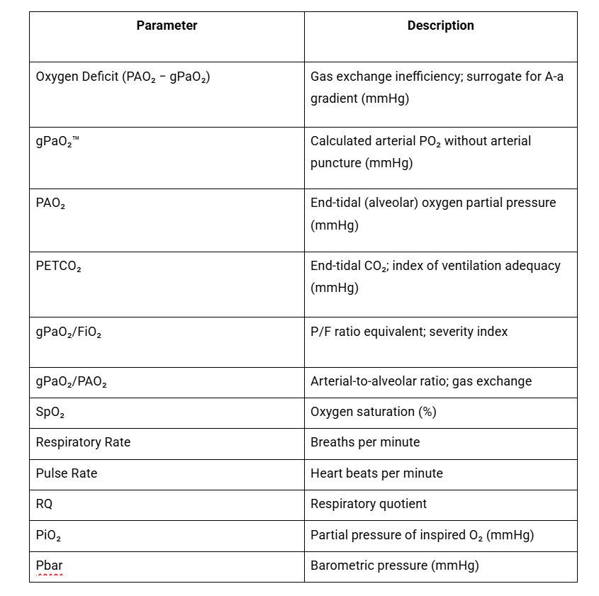
This multi-parameter profile gives the clinician a three-dimensional view: the alveolar gas composition (what the lung is presenting to the capillary bed), the arterial oxygen content (what the blood is actually receiving), and the efficiency of transfer between the two (where they diverge). This is information that neither SpO₂ alone, nor spirometry alone, nor symptom scores can provide.
Why This Is Particularly Relevant in the Indian Clinical Context
In India, the clinical setting where COPD management most often occurs is the outpatient chest clinic, not a fully equipped pulmonary function laboratory with trained technicians and calibrated multi-gas analyzers. The AGM100 requires only steady-state tidal breathing. No forced expiratory maneuver. No patient effort-dependence. No arterial puncture. Training requires less than one hour. The measurement takes minutes.
For a country where spirometry infrastructure is concentrated in urban tertiary centers, where first-line respiratory care is delivered by chest physicians operating in resource-constrained settings, and where patient populations include many who would struggle with the effort-dependent demands of high-quality spirometry, the operational profile of the AGM100 is a meaningful fit.
The Clinical Evidence: What the Peer-Reviewed Literature Shows
The scientific foundation of the AGM100 rests on a substantial body of published research. The evidence base spans physiological validation, disease-specific applications, and prospective clinical outcome studies.
Validation Against the Gold Standard: ABG-Level Accuracy Without the Needle
A landmark study by West et al., published in CHEST (2018), demonstrated that the Oxygen Deficit measured non-invasively correlated strongly with the A-a gradient calculated from simultaneously obtained arterial blood gases in outpatients with established cardiopulmonary disease. In patients with lung disease, the mean Oxygen Deficit was 48.7 ± 3.1 mmHg, in stark contrast to 4.0 ± 0.88 mmHg in healthy controls (p < 0.0001). The authors concluded that this non-invasive method “is very sensitive to the presence of disease and may discard the need for arterial punctures in many instances.”
The Ainslie et al. validation study, published in CHEST (2020), tested the AGM100 under conditions of hypoxic exercise, a physiological stressor that maximally taxes gas exchange reserves. Under rest and hypoxic exercise conditions, strong correlations between gPaO₂ and directly measured PaO₂ were found (R² = 0.97; mean bias = 1.70 mmHg). The authors concluded the AGM100 “provided a valid and reliable measure against directly measured arterial blood gases at rest and during hypoxic exercise.”
Validation Specifically in COPD Patients: The High-Altitude Study
Perhaps the most directly relevant study for Indian pulmonologists is the Champigneulle et al. validation study, published in J Clin Med (2023), which enrolled 131 moderate-to-severe COPD patients, with a mean FEV₁ of 60 ± 10% predicted, and measured concurrent AGM100 cPaO₂ against a reference portable arterial blood gas analyser. The study confirmed the AGM100’s accuracy in detecting severe resting hypoxemia at thresholds directly relevant to long-term oxygen therapy (LTOT) prescribing PaO₂ < 60 mmHg and ≤ 55 mmHg. This is a clinical decision that currently requires ABG measurement at most Indian hospitals, often creating a significant logistical burden for patients travelling from remote areas.
Predicting Respiratory Failure Before It Happens: The AUROC 0.94 Finding
An NIH-funded study published in J Clin Med (2025) evaluated the AGM100’s ability to predict imminent respiratory failure across a broad spectrum of cardiopulmonary conditions, including COPD, asthma, heart failure, and pulmonary embolism. Using need for supplemental oxygen within 24 hours as a clinically validated proxy for deterioration, the Oxygen Deficit predicted this outcome with an AUROC of 0.94 (p < 0.001), representing the highest reported prognostic accuracy for any non-invasive respiratory metric in the published literature.
An earlier ATS 2022 study provided even more striking data: an Oxygen Deficit of 40 mmHg or above predicted the need for supplemental oxygen during hospitalisation with an AUROC of 0.99, essentially a single, easily obtained non-invasive measurement providing near-perfect discriminatory accuracy. In India, where emergency respiratory triage in overloaded district hospitals or tertiary care centers is a daily clinical challenge, a non-invasive, instantaneous measurement with this level of discriminatory accuracy represents a meaningful addition to the clinical toolkit.
The V/Q Mismatch Data: Gas Exchange Impairment at GOLD Stage 1
Independent of the AGM100-specific studies, the fundamental physiology supporting gas exchange assessment in early COPD is well established. Rodriguez-Roisin and colleagues demonstrated using the multiple inert gas elimination technique (MIGET) that in patients at GOLD Stage 1, V/Q imbalance and the alveolar-arterial oxygen difference are already clearly abnormal, and that this gas exchange impairment is disproportionately greater than the airflow limitation at this stage. The clinical implication is profound: gas exchange assessment can detect physiologically significant COPD-related dysfunction at a stage when spirometry is just barely crossing the diagnostic threshold, or even before.
The Indian COPD Patient: Where Gas Exchange Assessment Adds the Most Incremental Value
The Indian clinical context creates specific scenarios where gas exchange assessment adds value that spirometry and pulse oximetry cannot provide. Let us look at a few illustrative scenarios.
Scenario 1: The Non-Smoking Rural Woman With Biomass Exposure
A 52-year-old woman from rural Maharashtra presents with progressive breathlessness and morning cough for three years. She is a non-smoker and has never been told she has lung disease. She has cooked over a wood fire in an unventilated kitchen for 28 years. Her resting SpO₂ is 95%. She does not have access to spirometry in her nearest primary health centre.
This patient represents one of the largest, most underserved COPD populations in India, and she is almost entirely invisible to existing diagnostic pathways. A non-invasive gas exchange measurement, requiring only steady-state tidal breathing, no arterial puncture, no special equipment infrastructure, could provide an immediate, objective assessment of whether her lungs are exchanging gas efficiently. An elevated Oxygen Deficit would confirm physiological impairment, justify further workup (spirometry at a referral centre, DLCO, imaging), and motivate intervention (fuel transition, pharmacotherapy, symptom management) without requiring her to make a lengthy journey for a laboratory-based test.
Scenario 2: The Post-TB Patient Cleared as Cured
A 44-year-old man from Jharkhand completed anti-tubercular therapy 14 months ago and was declared cured. He continues to experience exertional dyspnea and fatigue. His sputum is negative. His chest X-ray shows residual fibrotic changes in the right upper lobe. His resting SpO₂ is 96%. He has been reassured that his TB is cured and his symptoms are “normal sequelae.”
Post-tubercular COPD and post-tubercular gas exchange impairment are systematically underdiagnosed in India. Studies have found that 70% of post-TB patients have abnormal spirometry and 62.2% have reduced DLCO after completion of treatment, and that early identification and treatment of post-tubercular COPD meaningfully improves quality of life and reduces morbidity. An elevated Oxygen Deficit in this patient is objective physiological evidence that his lungs are not functioning normally, evidence that warrants investigation rather than dismissal, and that could initiate the referral and management pathway his condition requires.
Scenario 3: The Outpatient COPD Patient Between GOLD Stage 2 and 3
A 62-year-old retired government officer in Chennai, a former smoker, has confirmed COPD with spirometry from two years ago showing FEV₁ 58% predicted. He is on a LABA/LAMA combination. His latest spirometry at his follow-up visit shows FEV₁ 55% predicted, a 3% decline that falls within measurement variability. His symptom questionnaire (CAT score 16) is unchanged. His SpO₂ is 94%. Should his treatment be escalated? Is he progressing?
His AGM100 measurement reveals an Oxygen Deficit of 33 mmHg, elevated, and higher than the 22 mmHg recorded at his last visit. His PETCO₂ is at the low boundary of normal, suggesting compensatory hyperventilation. This gas exchange profile is the objective correlate of physiological worsening, even in the absence of spirometric progression that exceeds measurement variability. It changes the clinical decision: a structured conversation about escalating therapy, consideration of formal pulmonary rehabilitation, and closer monitoring on a 6-week rather than 6-month interval.
Scenario 4: The Acute Exacerbation Triage in a Busy Emergency Department
A 67-year-old known COPD patient arrives at the emergency department of a district hospital in Lucknow with increased breathlessness. SpO₂ is 91% on room air. The emergency physician must decide: observation ward or ICU? Supplemental oxygen titration or non-invasive ventilation? There is a queue of 40 patients.
A three-minute AGM100 measurement reveals an Oxygen Deficit of 52 mmHg with a gPaO₂ of 56 mmHg and low PETCO₂. This profile, significant V/Q mismatch, borderline arterial oxygenation, and compensatory hyperventilation, provides immediate, objective context for a triage decision that currently relies on clinical gestalt, SpO₂, and whatever ABG data can be obtained. The published evidence showing an AUROC of 0.94 for oxygen therapy prediction makes this non-invasive measurement a meaningful triage adjunct in precisely this high-stakes, time-pressured context.
The Limitations of Our Current COPD Monitoring in India
Indian pulmonologists managing COPD longitudinally rely on three primary assessment tools: spirometry, symptom questionnaires (CAT, mMRC), and exacerbation history. Each of these is valuable. Each has limitations that are particularly consequential in the Indian context.
Spirometry changes slowly, often imperceptibly from visit to visit, and requires patient effort-dependence that varies with operator skill and patient cooperation. In poorly characterized Indian populations (post-TB, biomass-related, occupational), standard reference equations developed on Western cohorts may systematically misclassify lung function severity.
Symptom questionnaires are subjective and influenced by cultural attitudes toward illness, health literacy, language barriers, and reporting bias. Indian patients, particularly rural and elderly patients with limited health literacy, may under-report symptoms, attributing progressive breathlessness to aging or physical deconditioning rather than lung disease.
Exacerbation history is retrospective and dependent on accurate recall and patient-reported outcomes. In Indian communities where every exacerbation requiring healthcare contact represents a significant financial and logistical burden, many exacerbations go unrecorded, either because the patient manages at home or because they access only informal or over-the-counter treatment.
The Oxygen Deficit adds something qualitatively different to this monitoring framework: a continuous, objective, quantitative, physiologically grounded index of gas exchange efficiency that changes in proportion to the patient’s real-time respiratory status. It is not effort-dependent. It is not language-dependent. It does not require patient recall. It measures what the lung is actually doing with oxygen, at the moment of assessment, without an arterial puncture.
A Vision for Indian Respiratory Practice: What Could Change
The diagnostic gap in Indian COPD care is not the result of inadequate clinical skill or physician motivation. It is the result of a structural mismatch between the scale of the disease, the heterogeneity of its etiological drivers, and the narrowness of the diagnostic toolkit that has been available.
Spirometry remains indispensable. DLCO is valuable. Symptom assessment is necessary. But COPD in India needs a complementary tool that:
– Works at the point of care, without laboratory infrastructure
– Does not require patient effort or technician skill
– Is not dependent on invasive procedures that carry cost, discomfort, and risk
– Is sensitive to physiological impairment earlier than spirometry
– Is relevant to the non-smoking, biomass-exposed, and post-tubercular COPD phenotypes that characterize so much of India’s disease
– Can be used for serial monitoring in routine outpatient practice
– Provides an objective, quantitative measure that goes beyond what pulse oximetry can offer
The MediPines AGM100 satisfies this specification with a level of clinical evidence, peer-reviewed, FDA-cleared, WHO-recognised, and CDSCO-approved that makes it a credible addition to the Indian respiratory clinician’s toolkit.
For the 37.8 million Indians currently estimated to have COPD, and the millions more whose disease remains entirely invisible to the healthcare system, a diagnostic evolution that begins detecting respiratory decline before symptoms fully declare themselves is not a luxury. It is a clinical necessity.
Conclusion: India Cannot Afford a Spirometry-Only Approach to COPD
The Indian COPD landscape is defined by three hard realities. First, the disease is vastly underdiagnosed, driven by structural barriers to spirometry access that are not close to being resolved. Second, India’s COPD population is phenotypically diverse in ways that spirometry-centric frameworks fail to capture, including the non-smoking biomass-exposed woman, the TB survivor, and the occupationally exposed laborer. Third, the disease causes disproportionate mortality in India precisely because it is detected late, managed inadequately, and monitored episodically.
Gas exchange assessment, operationalized through the FDA-cleared, WHO-recognized, peer-reviewed MediPines AGM100, offers Indian pulmonologists and chest physicians a physiologically grounded, clinically validated window into respiratory function that opens earlier, operates at the point of care, and provides information that spirometry and pulse oximetry simply cannot.
As the clinical evidence base grows, including from institutions and populations within India, the conversation about COPD assessment in India needs to move beyond the single question “does the patient have airflow obstruction?” to the richer question: “how efficiently is this patient’s lung exchanging oxygen, and is that efficiency declining?”
That question has an answer. The answer is now accessible, non-invasively, in minutes. The clinical imperative is to start asking it.
References
- GBD 2019 Chronic Respiratory Disease Collaborators. Indian Journal of Public Health. 2023. COPD in India: 37.8 million cases, 17.8% of global burden, 27.3% of global deaths.
- BMC Pulmonary Medicine. Burden of chronic obstructive pulmonary disease among Indian adults: systematic review and meta-analysis. 2026. Pooled prevalence 16% males, 9% females; 27% in elderly ≥60 years.
- Lung India. What is the true burden of chronic obstructive pulmonary disease in India? 2021;38(6):503-505. GBD 2019: COPD is 2nd leading cause of death and DALYs in India. COPD caused not only by tobacco but 48% of COPD deaths and 51% of COPD DALYs in India attributable to non-smoking causes (GBD 2019).
- PMC. Prevalence of COPD and determinants of underdiagnosis in women exposed to biomass fuel, India. 18.4% COPD confirmed in 2868 screened women; 14.2% newly diagnosed.
- PMC (Odisha study). Chronic bronchitis and airflow obstruction in never-smoking women with biomass fuel exposure. Airflow obstruction 22.4%; OR 5.55 for solid fuel use.
- PMC. Impact of biomass fuels on respiratory functions in rural India. India contributes 28% of deaths due to indoor air pollution from unprocessed fuels; 34,000 deaths/year in Indian women attributed to solid fuel COPD.
- PMC. Assessment of lung function in successfully treated tuberculosis in India. High burden of airflow obstruction and COPD in post-TB cases; screening recommended regardless of conventional COPD risk factors.
- PMC. Unveiling silent consequences: Impact of pulmonary tuberculosis on lung health. 70% abnormal spirometry, 62.2% reduced DLCO in post-TB patients.
- ERS. Under-diagnosis of COPD in primary care setting in Surat, India. 91.2% of COPD patients undiagnosed prior to study; GOLD Stage 2 (52.4%) and Stage 3 (39.2%) at time of first diagnosis.
- npj Primary Care Respiratory Medicine. Use of spirometry among chest physicians and primary care physicians in India. Family doctors and pediatricians rarely use spirometry; barriers include expense, uncertainty in interpretation, time constraints.
- Journal of Global Health Reports. Using spirometry for screening and diagnosis of chronic respiratory diseases in primary health care in rural India. 2024. Non-availability of spirometry cited as central barrier in rural settings.
- J Appl Physiol. Rodriguez-Roisin et al. V̇A/Q̇ imbalance and COPD staging severity. 2009. V̇A/Q̇ inequality disproportionately greater than airflow limitation at GOLD Stage 1.
- CHEST. West JB et al. A New, Noninvasive Method of Measuring Impaired Pulmonary Gas Exchange in Lung Disease: An Outpatient Study. 2018;154(2):363-369. Mean OD 48.7 ± 3.1 mmHg in lung disease vs 4.0 ± 0.88 mmHg in controls.
- CHEST. Ainslie PN et al. Validation of a Non-invasive Assessment of Pulmonary Gas Exchange During Exercise in Hypoxia. 2020. R² = 0.97; mean bias 1.70 mmHg.
- J Clin Med. Champigneulle B et al. Validation of Noninvasive Assessment of Pulmonary Gas Exchange in COPD during Initial Exposure to High Altitude. 2023;12(3):795. N=131 COPD patients; validated against portable ABG for LTOT thresholds.
- J Clin Med. McGuire WC et al. Noninvasive Assessment of Impaired Gas Exchange with the Alveolar Gas Monitor Predicts Clinical Deterioration in COVID-19 Patients. 2025;14(16):5880. AUROC 0.94, p<0.001; highest reported performance for any noninvasive respiratory metric.
- Medscape/ATS 2022. McGuire WC et al. Oxygen Deficit ≥40 mmHg predicted supplemental oxygen need with AUROC 0.99.
- WHO. 2021 Compendium of Innovative Health Technologies for Low-Resource Settings. MediPines AGM100 recognized.
- MediPines Corporation. Medicare APC Reimbursement. PRNewswire. August 6, 2025. CPT code 0893T operational.
- National Health Accounts, India 2023. Out-of-pocket health expenditure: 47.1% of total health expenditure in India.
Frequently Asked Questions About AGM100
How does the AGM100 measure alveolar oxygen (PAO₂)?
The AGM100 measures alveolar oxygen, expressed as end-tidal oxygen partial pressure (PAO₂), directly from the patient’s exhaled breath during normal, steady-state tidal breathing. As the patient breathes into the device, sensors capture the composition of exhaled gas at the end-tidal phase, the point in the breathing cycle when alveolar gas most closely represents the gas available at the alveolar-capillary interface. This end-tidal PAO₂ reflects the actual oxygen concentration that the lung is presenting to the pulmonary capillary blood for transfer. No forced manoeuvre, breath-hold, or arterial puncture is required. The measurement takes only a few minutes and updates in real time.
How does the AGM100 calculate the Oxygen Deficit?
The Oxygen Deficit is the AGM100’s primary index of gas exchange efficiency. It is calculated as the difference between the alveolar oxygen partial pressure (PAO₂) and the calculated arterial oxygen partial pressure (gPaO₂):
Oxygen Deficit = PAO₂ − gPaO₂
The device first measures PAO₂ directly from exhaled end-tidal gas. It then estimates gPaO₂, the arterial PaO₂, non-invasively, by combining the simultaneous pulse oximetry reading (SpO₂) with the measured end-tidal CO₂ (PETCO₂). The PETCO₂ is used to apply a Bohr effect correction to the oxyhemoglobin dissociation curve, enabling an accurate calculation of the partial pressure of oxygen in arterial blood from saturation data alone, without drawing blood. The gap between what the alveolus contains (PAO₂) and what the arterial blood receives (gPaO₂) is the Oxygen Deficit: a direct, quantitative measure of how efficiently, or inefficiently, the lung is transferring oxygen to the circulation.
In healthy adults at rest, the Oxygen Deficit is 0–15 mmHg. Any elevation beyond this range indicates gas exchange impairment. As Dr John B. West, Emeritus Professor of Medicine and Physiology at UC San Diego, states: “Oxygen Deficit is the most informative measure of pulmonary gas exchange.”
What is gPaO₂ and how does the AGM100 calculate it without an arterial blood gas?
gPaO₂ (calculated arterial partial pressure of oxygen) is the AGM100’s non-invasive estimate of the arterial PaO₂, the value that traditionally requires an arterial blood gas (ABG) sample. The device derives gPaO₂ by working backwards from the oxyhemoglobin dissociation curve. Since arterial oxygen saturation (SaO₂) and arterial PaO₂ have a well-defined mathematical relationship, and since that relationship shifts depending on the blood CO₂ level (the Bohr effect), the AGM100 uses the simultaneously measured PETCO₂ as a surrogate for blood CO₂ to correct the dissociation curve, then inverts it to calculate the PaO₂ that corresponds to the measured SpO₂. The result is a non-invasive, real-time estimate of arterial oxygenation that has been validated in peer-reviewed studies to show a near 1:1 relationship with directly measured ABG PaO₂.
How accurate is the AGM100 compared to arterial blood gas (ABG) analysis?
The AGM100 has been validated against arterial blood gas analysis in multiple peer-reviewed, published studies. The key evidence:
A landmark outpatient study by West et al., published in CHEST (2018), found that the Oxygen Deficit, as measured non-invasively by the AGM100, correlated strongly with the simultaneously measured A-a gradient from ABG in patients with established cardiopulmonary disease. In patients with lung disease, the mean Oxygen Deficit was 48.7 ± 3.1 mmHg vs. 4.0 ± 0.88 mmHg in healthy controls (p < 0.0001), confirming high sensitivity to disease.
The Ainslie et al. validation study, published in CHEST (2020), tested the AGM100 under hypoxic exercise — a condition that maximally stresses gas exchange. The study found an R² of 0.97 and a mean bias of just 1.70 mmHg between gPaO₂ and directly measured PaO₂, establishing a near 1:1 relationship. The authors concluded the AGM100 “provided a valid and reliable measure against directly measured arterial blood gases at rest and during hypoxic exercise.”
The Champigneulle et al. COPD-specific validation study (J Clin Med, 2023), enrolling 131 moderate-to-severe COPD patients, confirmed the AGM100’s accuracy in detecting hypoxemia at clinically relevant thresholds for long-term oxygen therapy prescribing.
This level of accuracy is what has enabled the AGM100 to receive FDA 510(k) clearance and MDSAP ISO 13485:2016 medical device certification, and what led the manufacturer to describe its precision as “comparable to arterial blood gas analysis.”
Can the AGM100 detect respiratory decline before symptoms appear in COPD?
Yes. And this is one of the most clinically significant applications of the device. In COPD, ventilation-perfusion (V/Q) mismatch and gas exchange impairment develop and worsen before airflow limitation becomes severe enough to produce prominent symptoms or cross the spirometric diagnostic threshold. A patient may maintain an SpO₂ of 94–96% and report manageable dyspnea, yet have a measurably elevated Oxygen Deficit, indicating that the lung is working harder than it should to maintain that saturation level and that its reserve capacity is already diminished.
Because the AGM100 measures the Oxygen Deficit at the point of care during normal breathing, not under laboratory-controlled conditions, it reflects the patient’s real-time physiological status. Serial measurements across outpatient visits can track a rising Oxygen Deficit over time, even when FEV₁ appears stable and symptoms remain unchanged. This rising trajectory is an objective early signal of worsening gas exchange, detectable weeks to months before clinical deterioration becomes evident. For COPD patients, particularly those with non-smoking risk factors such as biomass fuel exposure or post-tubercular lung disease, this early physiological signal is exactly the kind of information that can drive timely therapeutic intervention.
How does the AGM100 help in early detection of respiratory decline compared to pulse oximetry alone?
Pulse oximetry (SpO₂) is a valuable and ubiquitous tool, but it has a fundamental physiological limitation: SpO₂ does not fall meaningfully until arterial PaO₂ drops below approximately 60 mmHg, due to the flat upper portion of the oxyhemoglobin dissociation curve. A patient with significant V/Q mismatch and a widened alveolar-arterial oxygen gradient can maintain an SpO₂ of 95–96% through compensatory hyperventilation, appearing clinically stable, while the Oxygen Deficit is already substantially elevated.
The AGM100 closes this gap. By simultaneously measuring both what the alveolus contains (PAO₂) and what the arterial blood receives (gPaO₂), it quantifies the efficiency of oxygen transfer rather than just the end result of it. A rising Oxygen Deficit in the presence of a normal-appearing SpO₂ is an early physiological warning that the lung’s gas transfer mechanism is deteriorating, information that SpO₂ alone cannot provide. This makes the AGM100 a more sensitive early-warning tool for subclinical respiratory decline than pulse oximetry alone.
How does the AGM100 predict respiratory failure in COPD patients?
An NIH-funded, peer-reviewed study published in J Clin Med (2025) evaluated the AGM100’s ability to predict imminent respiratory failure across patients presenting with COPD, asthma, heart failure, and pulmonary embolism. Using need for supplemental oxygen within 24 hours as a validated real-world proxy for clinical deterioration, the study found that the Oxygen Deficit predicted this outcome with an AUROC of 0.94 (p < 0.001), the highest reported prognostic performance for any non-invasive respiratory metric in the published literature.
Earlier data presented at the American Thoracic Society (ATS) International Conference 2022 showed that an Oxygen Deficit of 40 mmHg or above predicted the need for supplemental oxygen at some point during the hospital course with an AUROC of 0.99, essentially a single, instantaneous, non-invasive measurement providing near-perfect discriminatory accuracy for oxygen therapy need. This makes the AGM100 a powerful triage tool in emergency and inpatient settings: a clinician can obtain an objective, evidence-backed assessment of a COPD patient’s risk of deterioration in minutes, without an arterial puncture.
Why do hospitals and clinics need the AGM100 if they already have ABG and spirometry?
ABG and spirometry are invaluable, but each has constraints that limit their use in everyday clinical decision-making:
ABG requires arterial puncture, is painful and carries procedural risk, demands laboratory processing time, is expensive to perform serially, and is logistically impractical for frequent monitoring, triage screening, or use outside of equipped facilities.
Spirometry measures airflow limitation, not gas exchange. It requires patient effort and cooperation, trained operators, and detects COPD only after significant structural damage has occurred. It cannot detect early gas exchange impairment and provides no real-time information about the severity of a patient’s current respiratory status.
The AGM100 fills the clinical gap between these two tools. It provides, non-invasively and at the point of care, the information about gas exchange efficiency that ABG offers, but in minutes, without a needle, and in any clinical setting. For hospitals, this means faster triage decisions, earlier identification of deteriorating patients, and objective data to guide oxygen therapy without waiting for ABG results. For outpatient clinics, it means visit-by-visit monitoring of COPD progression, treatment response assessment, and early detection of sub-clinical worsening, none of which spirometry or SpO₂ can accomplish with equivalent sensitivity or speed.
As one Milwaukee hospital’s Head of Respiratory described actual clinical practice: “We’re measuring these patients in the ER as a form of triage to determine what kind of care we’re going to provide. And then once the patient is admitted, we’re monitoring these patients at least twice a day to trend their responses to care and therapies. So far, it’s been extremely successful. We’re intervening much quicker.”

 By
By