Why Early Gas Exchange Assessment Matters in Clinical Practice
Overview
Hantavirus infection remains one of the most underrecognized yet clinically lethal zoonotic viral illnesses in modern medicine. Its significance lies in how rapidly a nonspecific febrile illness can transition into severe pulmonary compromise, often before traditional respiratory warning signs become apparent on conventional monitoring.
Hantavirus Pulmonary Syndrome (HPS) presents with fevers, myalgia, and severe respiratory compromise, with mortality rates reaching up to 40%. The most common etiological agents are the Sin Nombre virus in North America and the Andes virus in South America.
NCBI
For treating physicians, the challenge is not only diagnosis, but also recognising pulmonary physiological decline early enough to intervene effectively.
Understanding Hantavirus: A Zoonotic Disease with Serious Respiratory Consequences
Hantaviruses are spread through infected rodents via their urine, faeces, and saliva, with transmission occurring primarily through inhalation of the aerosolised virus.
Two major clinical syndromes are recognised:
1. Hemorrhagic Fever with Renal Syndrome (HFRS)
More prevalent in Europe and Asia, primarily involving renal and vascular injury. HFRS caused by Hantaan, Amur, and Dobrava viruses carries mortality rates of 5–15%, while Seoul virus causes moderate disease, and Puumala virus causes milder forms with mortality under 1%.
ScienceDirect
2. Hantavirus Pulmonary Syndrome (HPS) / Hantavirus Cardiopulmonary Syndrome (HCPS)
Predominant in the Americas, involving pulmonary and cardiovascular collapse. HCPS is geographically restricted to the Americas, where approximately 300 cases are diagnosed annually. In 2024, the United States reported 20 confirmed cases and 8 deaths, representing a 40% case fatality rate.
Medscape
Exposure Profile: Hantavirus Is Not an Occupational Disease Alone
A widely studied outbreak at Yosemite National Park demonstrated that hantavirus exposure is not confined to agricultural or occupational settings. Any exposure to rodent habitats, including cabin stays, camping, utility work, pest control, and farming, can increase the risk of disease.
Mayo Clinic
This makes a thorough exposure history critically important in the clinical evaluation of any unexplained febrile illness with respiratory progression.
Clinical Progression: Why Early Symptoms Are Misleading
Symptoms occur anywhere from one to eight weeks after exposure to the virus and unfold in three distinct phases. First is an early prodromal phase with flu-like symptoms, including fever, muscle aches, headache, and low platelet count. Second is the cardiopulmonary phase, during which patients may develop elevated or irregular heart rate, cardiogenic shock, and pulmonary capillary leakage leading to respiratory failure, hypotension, and fluid accumulation in the lungs and chest cavity. The final phase is recovery, which typically takes months, though breathing difficulties can persist for up to two years.
During the early phase, hantavirus can closely mimic influenza, dengue, leptospirosis, and viral pneumonia, making clinical differentiation particularly challenging.
Core Pathophysiology: HPS as a Gas Exchange and Endothelial Disorder
Hantavirus-associated respiratory failure is driven not by airway obstruction, but by targeted endothelial dysfunction at the pulmonary microvasculature.
Pathogenic hantaviruses selectively infect pulmonary microvascular endothelial cells, triggering innate and adaptive immune responses, including recruitment of CD8+ T cells and mononuclear effectors. This immune activation drives a massive release of proinflammatory cytokines, notably IL-6, TNF-α, and chemokine ligand 2, which disrupt vascular endothelial-cadherin, upregulate adhesion molecules, and force the formation of paracellular gaps that dramatically increase capillary permeability.
Activated macrophages and immunoblasts migrate to the interstitium of the lung. The subsequent secretion of TNF-α, IFN-γ, and nitric oxide results in pulmonary oedema and myocardial depression, potentially culminating in cardiogenic shock.
The functional consequences of this cascade are clinically profound: alveolar flooding, ventilation-perfusion (V/Q) mismatch, and impaired oxygen diffusion. Chest radiographs characteristically show peribronchial haze and Kerley’s B lines, which subsequently progress to alveolar flooding, manifesting as HCPS with concurrent circulatory shock and myocardial depression.
Critical Laboratory Red Flags
Laboratory findings often precede overt respiratory failure, making them a vital early warning system.
Progressive thrombocytopenia is one of the most consistent laboratory findings in HPS, occurring in virtually all patients and frequently present during the prodromal phase. Other important hematologic abnormalities include a left-shifted leukocytosis, presence of immunoblasts, and hemoconcentration, a tetrad seldom seen in other viral infections.
American Academy of Family Physicians
Thrombocytopenia, a left shift on peripheral smear, and an immunoblast count exceeding 10% of the total lymphoid series have been termed the diagnostic triad of HCPS. In experienced centres, this triad alerts practitioners to begin preparation for transfer to a facility capable of aggressive critical care management, including ECMO.
In a study of 567 suspected HPS patients, thrombocytopenia, suggestive chest X-ray findings, and receipt of supplemental oxygen demonstrated sensitivity above 95% for detecting HPS, while elevated hematocrit was 83% specific.
Why Traditional Monitoring May Miss Early Physiological Deterioration
Conventional bedside tools have meaningful limitations in capturing the early gas exchange dysfunction that precedes overt respiratory failure in HPS.
Pulse oximetry is a later marker; it may not capture evolving V/Q mismatch or subtle oxygen diffusion impairment until significant physiological compromise has already occurred.
Chest imaging provides structural information but not dynamic gas exchange data.
ABG testing is valuable but inherently episodic and invasive.
The clinical reality: a patient may appear relatively stable while their pulmonary gas exchange is already declining at the alveolar-capillary level. Patients are more likely to be receptive to oxygen therapy earlier in illness, making early diagnosis imperative. Initiating ECMO at the earliest sign of decompensation has an 80% survival rate despite cardiopulmonary collapse.
This underscores the critical value of detecting physiological deterioration before it becomes hemodynamically visible.
The Role of AGM100 in Early Gas Exchange Assessment
Because HPS is fundamentally characterized by evolving pulmonary gas exchange dysfunction driven by endothelial leakage and V/Q mismatch, tools that directly assess gas exchange physiology, rather than solely oxygen saturation, offer a clinically relevant adjunct in appropriate monitoring contexts.
The MediPines AGM100 evaluates oxygen deficit, estimated oxygenation impairment, pulmonary gas exchange abnormalities, and potential ventilation-perfusion disturbances on a non-invasive basis.
Potential clinical applications include:
1. Early Risk Stratification in Symptomatic Patients
In patients presenting with viral prodrome, rodent exposure history, mild dyspnea, or unexplained fatigue, AGM100 can help identify hidden physiological compromise earlier than symptom severity alone would suggest.
2. Serial Monitoring of Progression
Non-invasive gas exchange measurements may allow clinicians to track worsening oxygen deficit, progressive V/Q disturbance, and the need for escalation in respiratory evaluation.
3. Physiological Assessment Outside the ICU
Where physicians evaluate respiratory function in general wards or outpatient settings, AGM100 provides an additional physiological layer beyond pulse oximetry.
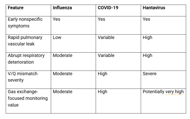
Comparative Context: HPS vs. Other Viral Pulmonary Syndromes
Key Clinical Red Flags: When to Suspect HPS
Consider hantavirus when the following factors coexist:
– Rodent or wilderness exposure history
– Fever with thrombocytopenia
– Severe myalgia disproportionate to the clinical picture
– Sudden or worsening dyspnea
– Bilateral pulmonary infiltrates on imaging
– Hemoconcentration (hematocrit >50% in men, >48% in women)
– Unexplained oxygenation decline without an alternative diagnosis
Important Clinical Clarification
AGM100 is not a diagnostic tool for hantavirus infection and does not replace RT-PCR, serology, CBC, imaging, or infectious disease evaluation. Its role is best understood as a non-invasive physiological monitoring adjunct that may help identify pulmonary gas exchange dysfunction earlier in appropriately selected patients, within approved clinical use settings.
Final Perspective
Because of the rarity of HPS and because of the need for early acute intervention in the face of precipitous decline, recognition of the unique laboratory profile of hantavirus pulmonary syndrome in the setting of a predisposing exposure history is of paramount importance.
Its trajectory is characteristically dangerous:
Viral prodrome → Hidden gas exchange impairment → Rapid pulmonary decline
In conditions where pulmonary dysfunction evolves faster than visible symptoms, earlier physiological assessment can improve clinical suspicion, monitoring precision, and escalation timing.
The AGM100 may offer physicians a valuable non-invasive adjunct in understanding gas exchange abnormalities sooner, supporting more informed respiratory evaluation in conditions where every hour of delay carries meaningful clinical consequences.
If you are a hospital or a healthcare provider, please write to us at info@stage021.com to learn more about the AGM100 and to schedule a live demo.

 By
By