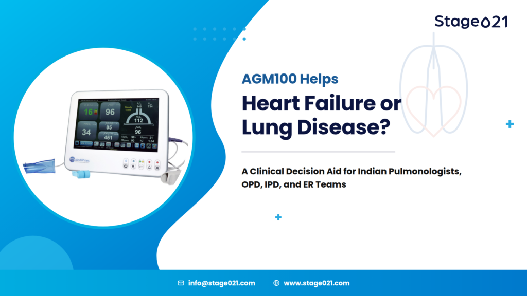
Heart Failure or Lung Disease? A Clinical Approach to Differentiating Dyspnea with AGM100
Breathlessness is one of the most common and most diagnostically ambiguous complaints seen across Indian outpatient departments, inpatient wards, and emergency rooms. It is also one of the few symptoms shared almost equally by cardiology and pulmonology: a patient in acute distress may be in decompensated heart failure, an acute exacerbation of COPD, or somewhere in the physiological overlap between the two. For the clinician at the bedside, the diagnostic question is rarely “is this patient breathless?”; it is “why.”
This article reviews the physiological overlap between cardiac and pulmonary causes of dyspnea, the limitations of standard bedside tools in resolving that overlap, and how non-invasive pulmonary gas exchange analysis, as performed by the AGM100 Gas Exchange Monitor, provides an additional, objective data point for clinicians managing breathless patients in OPD, IPD, and ER settings.
Heart Failure or Lung Disease? Why the Two Overlap
Dyspnea is not a diagnosis. It is a subjective symptom with an extensive differential; cardiac, pulmonary, mixed cardiopulmonary, metabolic, renal, and even psychogenic causes can all present similarly. Patients rarely describe it in physiological terms; instead, clinicians hear “breathing difficulty,” “chest tightness,” “air hunger,” or simply “I can’t catch my breath.”
The scale of the problem is well documented. Breathlessness is consistently reported as one of the leading reasons for emergency department presentation worldwide, and Indian data reflect the same pattern. A prospective study conducted in the emergency department of a large tertiary care teaching hospital in Vellore, Tamil Nadu, noted that acute dyspnea is among the most common reasons for ER visits, and that differentiating a cardiac from a pulmonary cause remains a persistent diagnostic challenge even with access to ultrasonography and standard investigations. This is not a gap unique to any one hospital or health system; it is a structural feature of how breathlessness presents.
Why the Heart and Lungs Are Diagnostically Inseparable
The cardiovascular and respiratory systems are physiologically coupled, not independent. The lungs oxygenate blood and clear carbon dioxide; the heart circulates that oxygenated blood to the rest of the body. A defect in either system can produce the same downstream symptom, breathlessness, through different mechanisms.
Primary pulmonary conditions such as COPD, bronchial asthma, pneumonia, and pulmonary fibrosis impair gas exchange at the level of the airway or alveolus, through airflow obstruction, ventilation-perfusion (V/Q) mismatch, or diffusion limitation.
Primary cardiac conditions, particularly congestive heart failure and pulmonary hypertension, impair gas exchange indirectly. As left ventricular function declines, pulmonary venous pressure rises, fluid accumulates in the interstitium, and the alveolar-capillary membrane thickens, producing a diffusion defect that looks physiologically similar to primary lung disease, even though the lungs themselves are structurally normal.
This is why respiratory symptoms in heart failure are so frequently mistaken for a primary pulmonary problem, and why COPD and heart failure are repeatedly described in the literature as the two most important differential diagnoses of dyspnea in older adults. Wheeze, orthopnea, exertional breathlessness, and nocturnal symptoms can appear in both conditions, sometimes in the same patient simultaneously. Cardiopulmonary comorbidity is common, not the exception.
Where Standard Bedside Tools Fall Short
Clinicians evaluating a breathless patient typically rely on history, physical examination, chest imaging, pulse oximetry, echocardiography, and pulmonary function testing. Each contributes meaningfully to the diagnostic picture, but none of them directly quantifies how efficiently oxygen is transferred from the alveolus into the pulmonary capillary in real time.
Pulse oximetry, in particular, is often treated as a proxy for respiratory status, but its limitations are increasingly well recognised in the literature. SpO₂ reflects oxygen saturation, not gas exchange efficiency; a patient can maintain a reassuring SpO₂ while significant ventilation-perfusion mismatch or diffusion impairment is already present. Peer-reviewed evaluations of pulse oximetry have documented discrepancies between SpO₂ and directly measured arterial saturation across a range of clinical scenarios, meaning a normal reading does not exclude clinically relevant gas exchange abnormality. For a symptom as physiologically ambiguous as dyspnea, this is a meaningful blind spot.
Arterial blood gas (ABG) analysis remains the reference standard for directly quantifying gas exchange, including the alveolar-arterial (A-a) oxygen gradient, the difference between alveolar and arterial oxygen tension, and a well-established parameter for narrowing the differential diagnosis of hypoxemia. But ABG is invasive, uncomfortable for the patient, and impractical to repeat serially through the course of a clinic visit or a hospital admission. In cardiology and pulmonology outpatient settings, repeated arterial puncture is rarely feasible.
This leaves a diagnostic gap: clinicians need objective, physiological information about gas exchange, but the reference-standard method for obtaining it is invasive and poorly suited to repeated, real-time assessment.
The Physiology Clinicians Are Really Trying to Capture
Understanding this gap requires returning to first principles. Gas exchange is the process by which oxygen moves from inspired air, through the alveoli, across the alveolar-capillary membrane, and into arterial blood, while carbon dioxide moves in the opposite direction to be exhaled.
The alveolar-arterial (A-a) gradient is the classic physiological marker of how well this process is working. In a healthy alveolar-capillary unit, alveolar and arterial oxygen tensions stay closely matched, and the A-a gradient remains narrow. When disease disrupts this interface, through obstruction, V/Q mismatch, diffusion limitation, or shunting, the gradient widens.
What makes the A-a gradient clinically useful is that it widens for both pulmonary and cardiac reasons. In COPD, it reflects airway obstruction and V/Q mismatch. In heart failure, published physiological studies describe an analogous widening driven by pulmonary interstitial congestion and reduced alveolar-capillary diffusing capacity, even in the absence of primary lung pathology. In other words, an abnormal gas exchange parameter does not, by itself, tell a clinician whether the origin is cardiac or pulmonary, but it does tell them that the alveolar-capillary interface is physiologically compromised, prompting the kind of structured workup (echocardiography, natriuretic peptides, spirometry, imaging) needed to localise the cause.
The AGM100 Gas Exchange Monitor is designed to bring this category of physiological information to the bedside without arterial sampling. Using a brief, non-invasive breathing manoeuvre, the device analyses exhaled gas concentrations alongside peripheral oxygen saturation to calculate a set of gas exchange parameters, including:
- Oxygen Deficit (OD): reflecting the gap between alveolar and estimated arterial oxygen, conceptually related to the A-a gradient
- gPaO₂™: a gas-based estimate of arterial oxygen tension, calculated without arterial puncture
- PAO₂: alveolar oxygen concentration, a foundational input for gas exchange assessment
- PETCO₂: end-tidal carbon dioxide, providing information on ventilation
Because the measurement is non-invasive and takes only a few minutes, it can be repeated across a clinic visit, an ER evaluation, or serially through an inpatient admission, something arterial sampling does not comfortably allow. This is particularly relevant for monitoring trends over time in patients with chronic cardiopulmonary disease, where a single snapshot is often less useful than the trajectory.
It is important to be precise about what this represents clinically: AGM100 is intended as an adjunct to, not a replacement for, existing diagnostic tools such as ABG, echocardiography, chest imaging, and pulmonary function testing. No single parameter, gas-based or otherwise, independently diagnoses heart failure or lung disease. What non-invasive gas exchange data adds is an objective, repeatable physiological signal that can be layered onto the existing clinical picture, particularly in patients where the cause of dyspnea is not yet clear.
Where AGM100 Fits Into Indian Clinical Practice
AGM100 is FDA-cleared and CDSCO-approved for use in India, and is intended for OPD, IPD, and ER settings. Within that scope, a few practical applications stand out for Indian pulmonologists, cardiologists, and emergency physicians:
- OPD triage of undifferentiated breathlessness: adding an objective gas exchange data point when history, examination, and SpO₂ alone leave the cardiac-versus-pulmonary question open
- Serial monitoring in IPD settings: tracking gas exchange trends through the course of treatment for heart failure, COPD exacerbation, or pneumonia, without repeated arterial puncture
- ER evaluation of acute dyspnea: supplementing pulse oximetry with a more complete picture of gas exchange efficiency during initial triage, particularly given the well-documented overlap between acute decompensated heart failure and acute respiratory illness in Indian ED presentations
- Outpatient follow-up of chronic cardiopulmonary disease: enabling repeatable, non-invasive assessment across visits, which is difficult to achieve with ABG alone
In each of these settings, the device is positioned to work alongside, not instead of, the diagnostic pathway Indian clinicians already use: clinical history, examination, chest imaging, echocardiography, natriuretic peptides where indicated, spirometry, and ABG when arterial sampling is clinically warranted.
Why Non-Invasive, Repeatable Gas Exchange Assessment Matters
Two structural realities make this category of tool particularly relevant to Indian healthcare delivery. First, resource and infrastructure constraints in many outpatient and ER settings make routine, serial ABG testing impractical, even when it would be clinically informative. Second, India’s burden of both cardiovascular and respiratory disease, including COPD linked to biomass and ambient air pollution exposure, alongside a substantial and growing burden of heart failure, means clinicians are frequently managing overlapping cardiopulmonary pathology in the same patient, not one condition in isolation.
A non-invasive tool that can be used repeatedly, without patient discomfort or the resource burden of arterial sampling, fits naturally into this environment, supporting earlier recognition of physiological deterioration, more informed triage decisions, and better-documented monitoring of chronic disease trajectory over time.
Conclusion
Differentiating cardiac from pulmonary dyspnea remains one of the more persistent challenges in day-to-day clinical practice, in India and internationally. Standard tools, history, examination, imaging, pulse oximetry, and echocardiography each contribute part of the picture, but none directly and repeatedly quantifies pulmonary gas exchange at the bedside without arterial sampling.
Non-invasive gas exchange analysis, as performed by AGM100, is designed to close part of that gap, offering clinicians an additional, objective, repeatable physiological signal to support the evaluation of breathless patients across OPD, IPD, and ER settings, alongside the diagnostic tools already in routine use.
If you are a hospital or healthcare provider and want to learn more about AGM100, please write to us at info@stage021.com or visit AGM100.
References
Chandy GM, Sathyendra S, Pichamuthu K, Hazra D, Abhilash KPP. Differentiating Cardiac and Pulmonary Causes of Dyspnea Using Ultrasonography and Dyspnea Discrimination Index. Indian J Crit Care Med. 2022;26(1):33–38. https://www.ijccm.org/abstractArticleContentBrowse/IJCCM/26431/JPJ/fullText
Shafi M. Differentiating Cardiac and Pulmonary Causes of Dyspnea: Is Point-of-care Ultrasound the Ultimate Tool? Indian J Crit Care Med. 2022;26(1):7–8. https://pmc.ncbi.nlm.nih.gov/articles/PMC8783231/
COPD and heart failure: differential diagnosis and comorbidity. PubMed. https://pubmed.ncbi.nlm.nih.gov/31111162/
Pulse Oximetry: Uses and Limitations. The Journal for Nurse Practitioners. https://www.npjournal.org/article/S1555-4155(07)00210-3/fulltext
Impacts of Skin Colour and Hypoxemia on Noninvasive Assessment of Peripheral Blood Oxygen Saturation: A Scoping Review. PMC. https://www.ncbi.nlm.nih.gov/pmc/articles/PMC10610303/
Physiology, Alveolar to Arterial Oxygen Gradient. StatPearls, NCBI Bookshelf. https://www.ncbi.nlm.nih.gov/books/NBK545153/
Diagnostic Evaluation of Dyspnea. American Family Physician. https://www.aafp.org/pubs/afp/issues/1998/0215/p711.html
Morosin M, Vignati C, Novi A, et al. The alveolar-to-arterial oxygen partial pressure difference is associated with pulmonary diffusing capacity in patients with heart failure. Respir Physiol Neurobiol. 2016;233:1-6. https://pubmed.ncbi.nlm.nih.gov/27374970/
The Alveolar Gas Monitor: An Alternative to Pulse Oximetry for the Noninvasive Assessment of Impaired Gas Exchange in Patients at Risk of Respiratory Deterioration. PMC. https://www.ncbi.nlm.nih.gov/pmc/articles/PMC12387870/
Noninvasive Assessment of Impaired Gas Exchange with the Alveolar Gas Monitor Predicts Clinical Deterioration in COVID-19 Patients. PMC. https://www.ncbi.nlm.nih.gov/pmc/articles/PMC10573776/
This content is intended for healthcare professionals and is provided for educational purposes. It does not constitute medical advice or a substitute for clinical judgment.
Frequently Asked Questions About AGM100
Is AGM100 a replacement for arterial blood gas (ABG) testing?
No. AGM100 is intended as a non-invasive adjunct to existing diagnostic tools, including ABG, not a replacement for it. ABG remains the reference standard for direct arterial gas measurement, particularly when arterial sampling is clinically indicated.
Can AGM100 tell a clinician whether a patient has heart failure or lung disease?
Not on its own. Gas exchange parameters such as Oxygen Deficit reflect how efficiently oxygen is transferred across the alveolar-capillary membrane, but an abnormal result can arise from either cardiac or pulmonary pathology. It is intended to be interpreted alongside history, examination, imaging, echocardiography, and other standard investigations.
Where does AGM100 fit within the existing diagnostic pathway for breathlessness?
AGM100 is used alongside, not instead of, the tools already in routine use for evaluating dyspnea: clinical history and examination, chest imaging, echocardiography, natriuretic peptides where indicated, spirometry, and ABG when arterial sampling is clinically warranted. It adds a non-invasive, repeatable gas exchange data point to that existing workflow, intended for use across OPD, IPD, and ER settings.
How is the AGM100 test performed?
The patient performs a brief, non-invasive breathing manoeuvre into the device. The system analyses exhaled gas concentrations alongside peripheral oxygen saturation to calculate parameters including Oxygen Deficit, gPaO₂™, PAO₂, and PETCO₂, typically within a few minutes.
How does AGM100 compare to spirometry or pulmonary function testing?
Spirometry and pulmonary function testing assess airflow and lung mechanics, how well the lungs move air in and out. AGM100 assesses a different physiological dimension: gas exchange efficiency, or how effectively oxygen moves from the alveoli into the bloodstream. The two are complementary rather than interchangeable, and are often useful together when characterising a patient’s respiratory status.
How does AGM100 fit into existing hospital or clinic workflows?
AGM100 is designed to slot into existing OPD, IPD, and ER workflows as a bedside or in-clinic assessment, rather than requiring a dedicated lab or specialist referral. Results are generated on the device within a few minutes, allowing clinicians to incorporate gas exchange data into the same visit or encounter as history, examination, and other point-of-care findings.
Can AGM100 results be documented, printed, or stored for patient records?
Yes. The device generates a results summary that can be documented as part of the patient record, supporting both point-in-time assessment and trend tracking across repeat visits or an inpatient stay.
How often can a patient be tested on AGM100?
Because the assessment is non-invasive and does not involve arterial puncture, it can be repeated as clinically indicated, for example, to track gas exchange trends through the course of treatment for heart failure or a COPD exacerbation, or at follow-up visits for chronic respiratory or cardiac disease.
How is AGM100 different from a standard pulse oximeter?
A pulse oximeter estimates oxygen saturation alone. AGM100 evaluates broader gas exchange physiology, including Oxygen Deficit, gPaO₂™, PAO₂, and PETCO₂, offering clinicians a more complete, though still adjunctive, picture of pulmonary gas exchange efficiency.
How long does an AGM100 assessment take, and what does it involve for the patient?
The test involves a brief, non-invasive breathing manoeuvre performed at the bedside or in-clinic, with results typically available within 2 minutes. No arterial puncture, sedation, or special patient preparation is required.

 By
By